Microphotography contest 2026
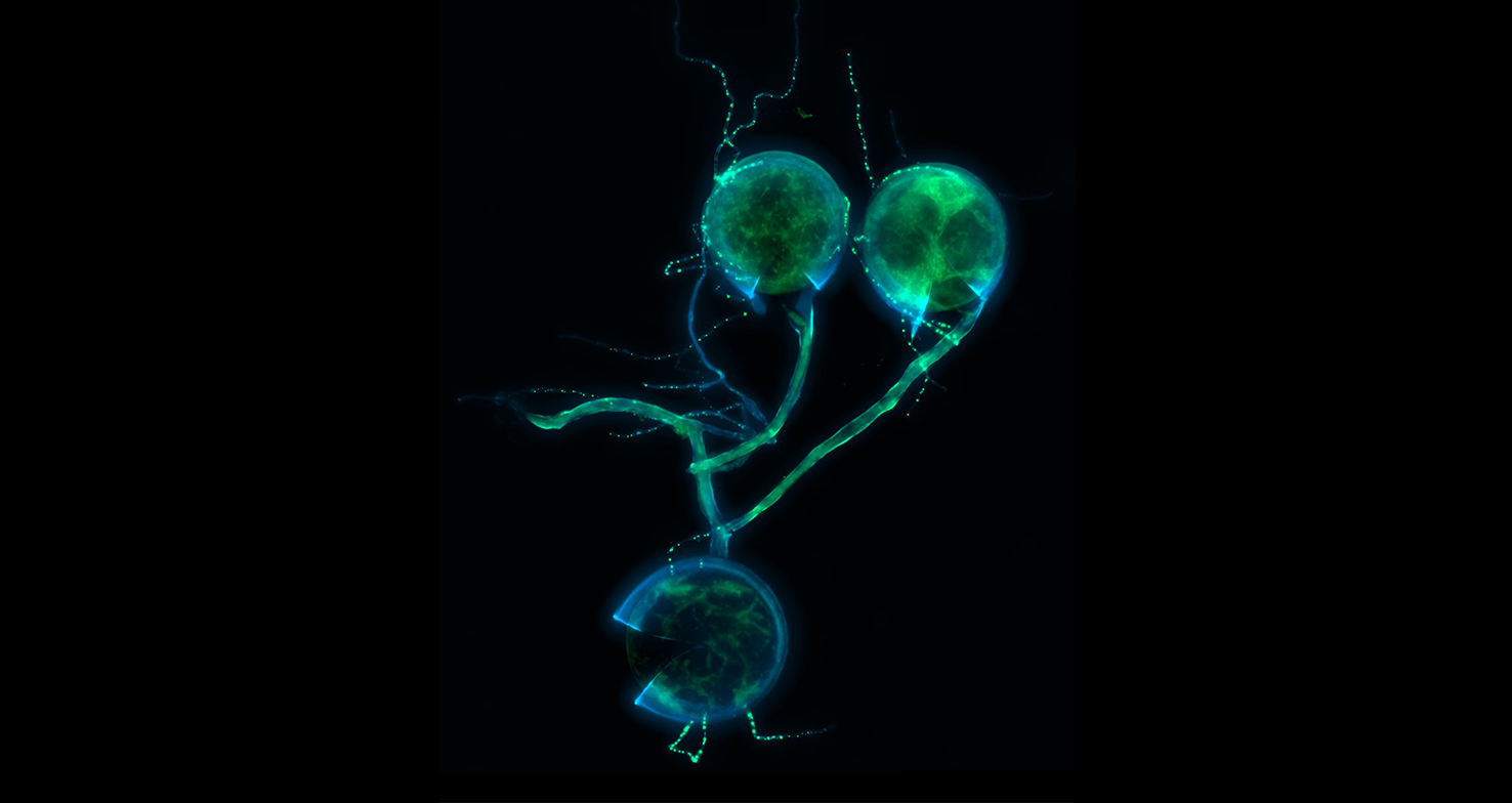
The winner of the 2026 KNVM Microphotograph competition is:
"Spores - inner and outer space" by Midge Woodward (VU Amsterdam)
Shortlist of selected images in 2026:
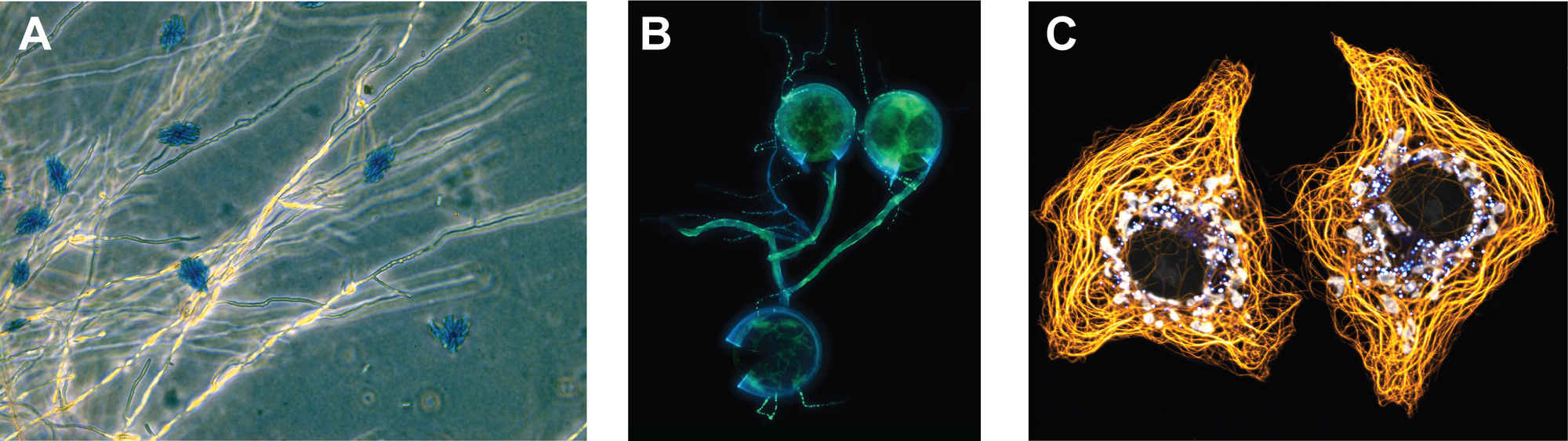
A | Schizophyllum commune & Indigotin | Alarik van Diepeningen - WUR
Several of my Schizophyllum commune lineages spontaneously started
to produce indigotin, a bright blue pigment that is insoluble in water and
crystalizes.
B | Spores - inner and outer space | Midge Woodward - VU Amsterdam
Three germinating spores of the arbuscular mycorrhizal fungus Rhizophagus irregularis,
co-stained with fluorescent dyes Calcofluor White (chitin, in blue) and SYTO-13 (nucleic
acids, in green). Playing with incubation times, I caught the spores before the nucleic acid
dye had time to bind all nuclei and mitochondria. The unbound dye reveals other structural
features of the fungus’ interior; a wrinkling membrane, perhaps gaps between lipids, the
cytoplasm of connecting hyphae. Sometimes unintentional or mistaken images can reveal
more about the character of an organism than those we intend to capture for data collection.
I am always trying to peer closer and closer into the fungal cell, but the underground and
its insides often feel further away and more mysterious than the planets and stars.
C | Golden Cytoskeleton & Lipid Gems | Walid Idi - Université Laval, Canada
Confocal microscopy image showing COS-7 cells highlighting the intimate
interplay between metabolic organelles. Lipid droplets are stained with
LipidSpot (in brilliant blue), while mitochondria are visualized using an
anti-Tom20 antibody (in white). The cytoskeletal architecture is revealed
by an anti-alpha-tubulin antibody (in gold), offering a detailed view of the
microtubule network and its spatial relationship with mitochondria and
lipid droplets.